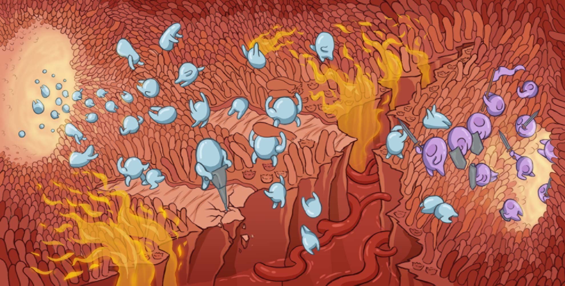
We are studying how the intestinal epithelium combats enteric pathogens. We believe that to gain a comprehensive understanding of the molecular mechanisms regulating gut homeostasis (finely tuned balance between response against enteric pathogens vs. tolerance of commensals), an integrative system approach needs to be implemented and exploited. To achieve this, we combine stem cell biology, virology, genomics, bioengineering, and cell biology to dissect host/pathogen interactions at the intestinal mucosa surface. Our long-term goal is to develop technologies and cross disciplinary pipelines to tailor pharmacological interventions to both the host (patient specific, cell type specific or pathology specific) and the pathogen (norovirus, coronavirus, rotavirus, astrovirus).
Not all intestinal epithelial cells are the same. They are compartmentalized through the head-codal axis facing different microenvironments depending on their location (e.g. duodenum, jejunum, ileum, and colon) providing them with different and unique functions. Additionally, intestinal epithelium cells are organized in a very specific manner throughout the crypt-villi axis facing different concentrations of oxygen. In this research axis, we are developing methods to integrate the gut specific microenvironment (e.g. low oxygen or hypoxia) to evaluate how it impacts the metabolic state of intestinal epithelial cells, their differentiation status and their response against pathogens and commensals. We compare how these mechanisms differ between the different GI sections (e.g. duodenum, jejunum, ileum, and colon) to understand why pathogens have section-specific tropism.
The intestinal epithelium is composed of multiple cell types that are all derived from a unique stem cell niche. In this research axis, we are comparing how the different cell types respond to the same enteric pathogen challenge. We use a combination of organoid-model systems, high spatial-temporal resolution live microscopy, single cell transcriptomic approaches, and spatial transcriptomics of tissue to decipher how stem cells are rewired to create different intestinal lineages (e.g. enterocytes, goblet cells, Paneth cells, etc) that use different strategies to combat enteric viruses (e.g. different pathogen recognition receptors, different response to interferon, expression of different interferon stimulated genes (ISGs) etc). This characterization will allow us to better understand enteric pathogen tropism and cell type specific pharmacological interventions to combat infections or to regulate inflammatory bowel disease.
Intestinal epithelial cells are polarized. They have an apical side which faces the lumen of the gut and a basolateral side facing the lamina propria. The apical and basolateral membranes have a distinct lipid and protein composition providing epithelial cells their unique function at a mucosal surface. While we have previously shown that polarity is key to regulate host/pathogen interactions in the human gut, it remains unclear how polarity is established and maintained at the molecular level. Similarly, it remains unknown whether these mechanisms are similar or distinct between the different intestinal lineages. In this research axis, we employ proteomic proximity assays and CRISPR knock-in technologies to refine our molecular understanding of cellular polarity.
We use multi-color fluorescent in-situ hybridization (FISH) to evaluate virus infection and immune response at the single cell level.
We employ live-cell microscopy to track virus infection and spread and the corresponding induction of immune response in our epithelial models.
We use single cell sequencing to unravel cell specific responses to viral infection in our organoid models.
We use patient derived organoids to model human organs with increasing complexity.